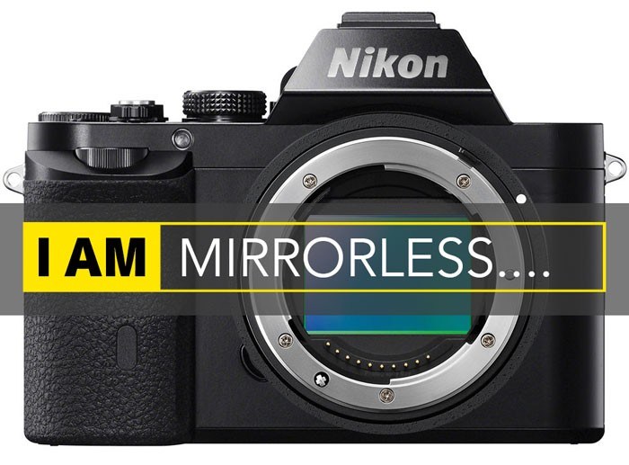

Recently, high-end digital single light reflex (DSLR's) cameras have been developed, which fulfill these criteria and are reasonably priced (approximately USD 2500) compared to HD medical cameras.

Only the high-end 3-chip CCD medical cameras (priced nearly USD 21,000) fulfilled these exacting requirements till now. Hence, vitreoretinal surgery recording needs a camera sensor with three qualities: (1) high quantum efficiency so that it can capture videos in low light, (2) high signal to noise ratio so that these images do not have grain, and (3) broad dynamic range so that both well-illuminated and dark areas are visualized equally. A bright light emitting diode light from the microscope is used for anterior segment surgeries while retinal surgeries are essentially performed in low light using the light from a fine endoilluminator probe. This is because of the difference in illumination sources used during surgery. Although these cameras give the acceptable quality video for recording anterior segment surgery, we observed that they are not suited for recording vitreoretinal surgeries. To overcome this, we explored various cost-effective alternatives such as single chip charged coupling device (CCD) and webcam-based cameras.

However, cost constraints come in the way of procuring these quality high definition (HD) recording systems. Numerous companies such as Sony, Ikegami, and Optronics manufacture high quality recording systems. Video-assisted skill transfer is very effective way to train residents during ophthalmic training.


 0 kommentar(er)
